Photoacoustic Neuroimaging with the TriTomTM
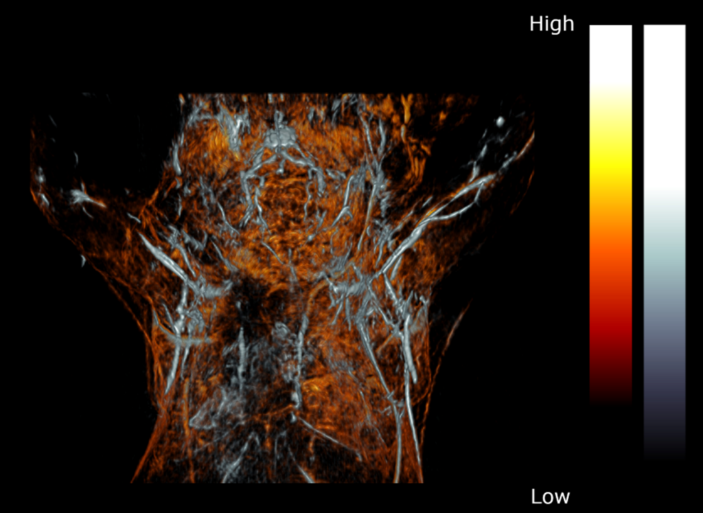
Brain imaging, or neuroimaging, is used to understand the relationship between brain function and behavior and study the underlying causes of neurological and psychiatric diseases. It is important in helping to understand the relationship between specific areas of the brain and what function they serve. Photoacoustic Imaging (PAI) relies on differential thermoelastic expansion. A pulsed laser light is used to illuminate a tissue of interest where the light is absorbed resulting in local heating and thermoelastic expansion. The produced pressure, or sound waves, are emitted and detected by high-frequency transducers allowing the signals to be processed into high-resolution images. Therefore, PAI combines the temporal and spatial resolution of ultrasound with the contrast and spectral nature of optics PAI is a noninvasive technique that can be used for multiple applications including brain imaging. It allows for an image of the brain with
micrometer-millisecond spatiotemporal resolution with sufficient penetration enabling the structural interrogation of the brain ranging from microscopic to macroscopic.
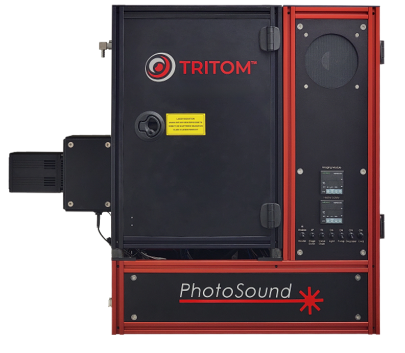
Imaging System TriTomTM
Repetition Rate 20 Hz
PA Excitation Range 532 nm & 650-1300 (2300) nm
Photoacoustic Neuroimaging
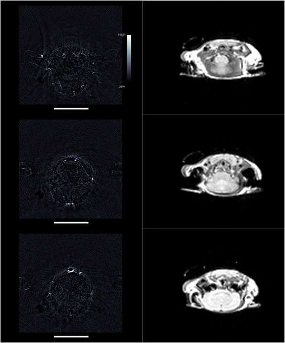
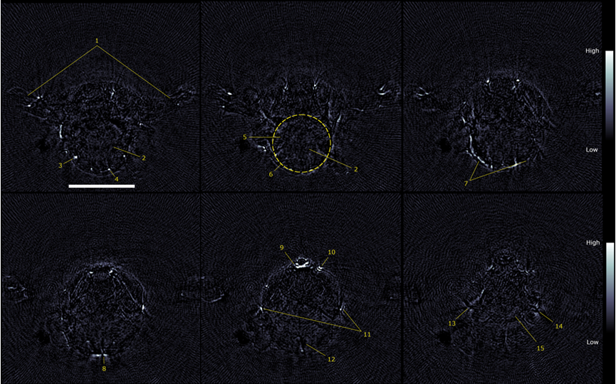
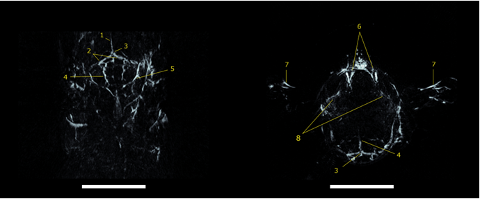
Neuroimaging, or brain imaging, is a fundamental tool for evaluating and monitoring the structure and function of the brain. However, there is currently no single imaging method capable of accurately mapping the brain’s complex anatomy and physiologic processes. Photoacoustic tomography (PAT) is a noninvasive and non-ionizing imaging modality that can be used as a complementary tool to conventional imaging techniques used in preclinical neuroscience research such as magnetic resonance imaging (MRI), computed tomography (CT), and positron emission tomography (PET). PAT is capable of acquiring high contrast and resolution images of optical absorption with great depths. PAT relies on the detection of acoustic ultrasound waves generated following the optical absorption of laser light by chromophores, i.e., hemoglobin, in biologic tissue. A tissue’s properties are dependent on the tissue’s constituents such as water, fat, melanin, oxy-hemoglobin, and deoxy-hemoglobin. The combination of optical excitation with ultrasonic detection of PAI can provide complementary information to other modalities and offers many advantages such as providing portable technology and no harmful ionizing radiation. PAT can provide high-resolution, high-sensitivity images with or without exogenous contrast.
PAT and MRI: Complimentary Imaging Modalities for Brain Imaging
MRI produces detailed images of organs and structures of the body by using large magnetic and radio waves and can acquire high-resolution images of brain structures over large volumes. However, it provides poor temporal resolution and it cannot be used to study fast hemodynamic responses and mechanisms. Optical imaging provides contrast for structural and functional imaging by exploiting diverse biological molecules present in tissues, but the optical scattering of the imaging technique limits the imaging depth. Utilizing the imaging capability of single or multiple components of noninvasive imaging tools, PAT and MRI can bridge the limitations of conventional brain imaging techniques by compensating the limitations of each imaging modality.