TriTom™ Photoacoustic Imaging Platform: Small Animal Anatomical Imaging
The ability to directly visualize and evaluate anatomical structures and biological processes in small animal models at the whole-body level is critical for understanding the pathophysiology of human diseases. The TriTom™ small animal imaging platform provides noninvasive high-resolution photoacoustic tomography (PAT) images that can be used to extract functional and anatomical information at the molecular level.
Whole Body Anatomical Imaging
Noninvasive whole-body imaging of small animals is crucial for understanding the fundamental relationship between anatomical structure and function. Optical methods have a high spatial resolution but suffer from shallow penetration depth (1-2 mm). Non-optical techniques such as MRI and PET provide the penetration depth but are costly, have long acquisition times, use ionizing radiation, or require exogenous contrast agents. Photoacoustic imaging takes advantage of the intrinsic optical properties of tissue, specifically hemoglobin, to provide high-resolution images of both superficial and deep blood-rich structures in murine models.
Imaging System TriTomTM
Repetition Rate 20 Hz
PA Excitation Range 532 nm & 650-1300
(2300) nm
Functional Imaging
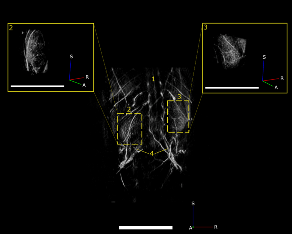
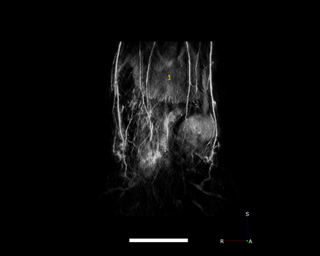
In addition to whole-body anatomical imaging, photoacoustic imaging can be utilized for functional imaging of physiological processes, which can include monitoring blood oxygenation, tumor growth, or therapeutic effects. Contrast agents, such as ICG, nanoparticles, etc., can be used to evaluate either blood-flow dynamics or for targeted imaging of vascular biomarkers. Here, we show high-resolution images of vasculature in the kidneys (Figure 2) and liver (Figure 3), demonstrating the ability to detect pathophysiological abnormalities in superficial and deep tissues.